Book a Home Collection Now!
How Is a Stroke Diagnosed and What Tests Are Required?
October 31, 2026 - By Lupin Diagnostics
Every minute matters when a stroke strikes. Understanding how a stroke is diagnosed can help you recognise why emergency teams move so quickly. A stroke cuts off blood supply to part of the brain, and rapid testing determines the type and guides treatment. This article walks you through the key assessments, imaging scans, blood tests, and cardiac diagnostics that doctors rely on to make a stroke diagnosis.
How Is a Stroke Diagnosed upon Emergency Arrival?
When someone arrives at the emergency room with suspected stroke symptoms, doctors begin a rapid, structured assessment. The goal is simple: confirm the stroke, identify its type, and act fast.
The medical team typically follows these steps:
- Neurological scoring: The National Institutes of Health Stroke Scale (NIHSS) is the most widely recommended tool for evaluating stroke patients in hospitals. It checks speech, vision, movement, and sensation.
- Medical history review: Doctors ask about symptom onset, existing conditions such as diabetes or hypertension, and current medications.
- Physical examination: Heart rate, blood pressure, and oxygen levels are checked immediately.
Up to 85% of strokes are ischaemic — caused by a blocked blood vessel. The remaining cases involve bleeding in the brain. Telling these two apart is critical because treatments differ entirely.
Also Read: Blood Clotting Disorders: Types, Symptoms & Treatment
What Tests Are Required to Diagnose Stroke Through Imaging?
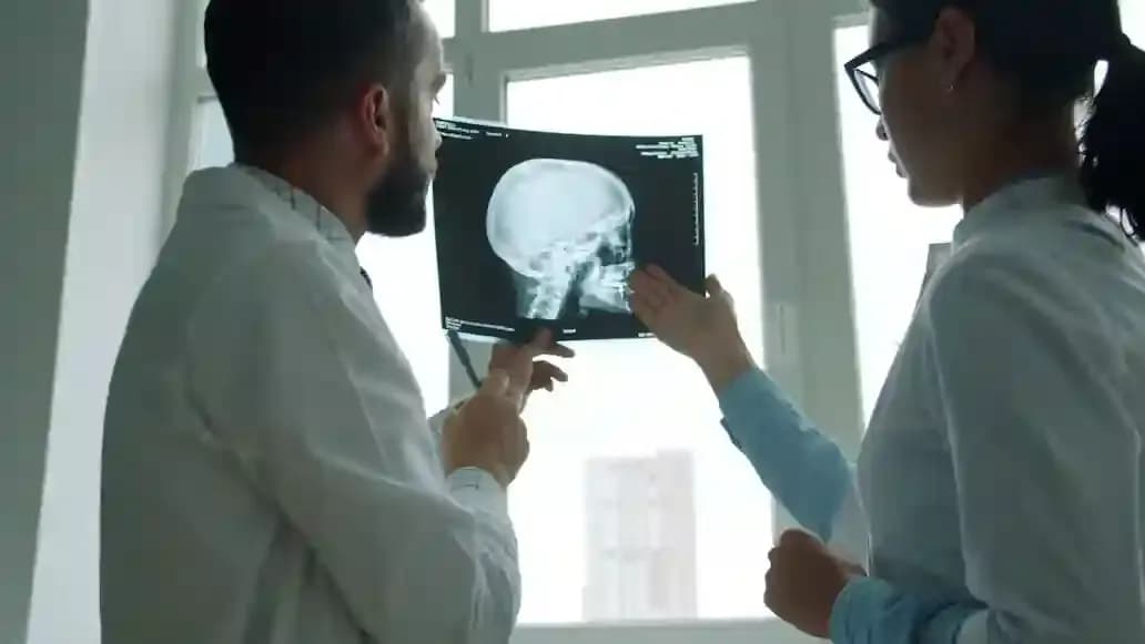
Brain imaging is the cornerstone of stroke diagnosis. The WHO recommends immediate imaging, CT or MRI, as soon as a stroke is suspected.
CT Scan
A non-contrast CT scan is usually the first test performed. It uses X-rays to create images of the brain within minutes. Its main role is ruling out bleeding. The scan is painless and widely available.
MRI With Diffusion-Weighted Imaging
MRI provides far more detailed brain images using magnetism. Diffusion-weighted imaging (DWI) is the most sensitive method for spotting early ischaemic damage. However, it takes around 30 minutes and is less available in smaller hospitals.
CT Angiography vs. MR Angiography
These specialised scans focus on blood vessels in the neck and brain. They help locate blockages or aneurysms that caused the stroke.
| Feature | CT Scan | MRI (DWI) |
|---|---|---|
| Speed | A few minutes | ~30 minutes |
| Availability | Widely available | Limited in smaller centres |
| Best For | Detecting bleeding | Detecting early ischaemic damage |
| Sensitivity for Ischaemia | Lower in early hours | Highest available |
Which Blood Tests Help During Stroke Evaluation?
Blood tests alone cannot confirm a stroke. However, they play a supporting role by ruling out conditions that mimic stroke symptoms. Your doctor will decide which tests are needed based on your specific situation.
Key blood tests typically ordered include:
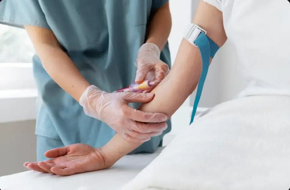
- Fasting Blood Sugar Test: Low blood sugar can cause symptoms that look like a stroke, making a Fasting Blood Sugar test a priority to rule this out.
- Complete Blood Count (CBC): Checks for infection, anaemia, or abnormal platelet counts through a complete blood count test.
- Coagulation tests (PT/INR, aPTT): These measure how quickly your blood forms a clot using PT/INR test and aPTT tests. They guide decisions about clot-dissolving treatments.
- Cardiac biomarkers: Troponin levels help check for heart-related complications.
These results, combined with imaging, give doctors a complete picture for planning treatment.
How Do Heart and Vessel Tests Identify Stroke Causes?
Once the immediate crisis is managed, doctors investigate the underlying cause. Heart and blood vessel tests are essential here.
- Carotid Doppler ultrasound: This non-invasive scan checks for plaque buildup or narrowing in the neck arteries that supply blood to the brain.
- Echocardiogram (Echo): An ultrasound of the heart that detects blood clots, structural defects, or other issues that may have caused a clot to travel to the brain.
- ECG and Holter monitoring: An electrocardiogram records heart rhythm to spot atrial fibrillation, an irregular heartbeat that significantly raises stroke risk. Longer monitoring catches irregular rhythms that brief tests might miss.
Also Read: Symptoms Of High Lipoprotein: Warning Signs & Heart Risks
What Happens After a Stroke Is Diagnosed?
Diagnostic results directly shape the emergency treatment plan. For ischaemic strokes, clot-dissolving medications such as alteplase or tenecteplase may be administered within a 4.5-hour window from symptom onset. Treatment decisions are always made by the medical team based on individual assessment.
For larger blockages, a procedure called endovascular thrombectomy may be considered. Extended treatment windows, up to 24 hours for thrombectomy, are sometimes possible based on advanced imaging results. Haemorrhagic strokes may require surgical intervention instead.
This is exactly why "time is brain." The faster the diagnosis, the more treatment options remain available.
Conclusion
Stroke diagnosis depends on a chain of rapid, coordinated tests. From the first CT scan in the emergency room to heart monitoring days later, each test adds a vital piece to the puzzle. If you or a loved one has risk factors like high blood pressure or diabetes, regular health check-ups can help catch warning signs early. Lupin Diagnostics offers comprehensive health check-up packages at NABL-accredited labs across India, a simple step towards staying ahead.
Frequently Asked Questions
How fast can a CT scan detect a stroke?
A CT scan takes only a few minutes and can quickly identify bleeding in the brain. Early ischaemic changes, however, may appear subtle on CT and sometimes require an MRI for confirmation.
Can a stroke show up on a blood test?
Blood tests cannot diagnose a stroke on their own. They help rule out conditions like low blood sugar or clotting disorders that can mimic or complicate stroke symptoms.
What happens if an MRI doesn't show a stroke?
This is not uncommon; up to 16% of acute ischaemic strokes may not appear on diffusion-weighted MRI. Doctors then rely on clinical symptoms and may repeat imaging later.
Is a lumbar puncture ever needed for stroke?
A lumbar puncture may be performed if a specific type of bleeding called subarachnoid haemorrhage is suspected but not visible on brain imaging.
How do doctors tell a stroke from a TIA?
A transient ischaemic attack (TIA) causes temporary symptoms without lasting brain damage on imaging. A stroke shows evidence of actual tissue injury on scans.
Can an EKG detect a stroke in progress?
An ECG cannot directly detect a stroke. It identifies irregular heart rhythms like atrial fibrillation, which is a common underlying cause of strokes.
This article is for informational and educational purposes only. It is not a substitute for professional medical advice, diagnosis, or treatment. If you suspect a stroke or have any health concerns, please seek immediate medical attention from a qualified healthcare provider.
Featured Articles

Dehydrated How To Make A Lifesaving Diy Rehydration Drink For Cholera Recovery
Posted on: 22nd Jan 2026
Diabetes Screening For Diagnosis And Prevention
Posted on: 25th Oct 2023

Eating To Recover: The Best Foods For Getting Back On Your Feet After Cholera
Posted on: 22nd Jan 2026