Histopathology Radical Resection Specimen Test: Purpose, Procedure, Results & Price
About Histopathology Radical Resection Specimen Test: Purpose, Procedure, Results & Price
| Field | Value |
|---|---|
| Also Known As | Histopathology – Large Complex / Cancer Specimen, Radical Specimen Test, Biopsy Material Radical, Histopathology Large Specimen (with lymph nodes), Surgical Pathology – Radical Resection |
| Sample Type | Surgically resected tissue specimen (organ or tumour with surrounding tissue and lymph nodes) |
| Fasting Required | No fasting required for the laboratory analysis; fasting before the surgical procedure may be directed by your surgeon |
| Report Time | 5 to 10 working days; may extend to 7 to 14 days if immunohistochemistry or special stains are needed |
| Recommended For | All genders and ages; primarily patients undergoing cancer surgery requiring tumour removal with margin and lymph node assessment |
| Price | Starting at ₹3,000 |
What is a Histopathology - Radical Resection Specimen (Histopath (Radical)) Test?
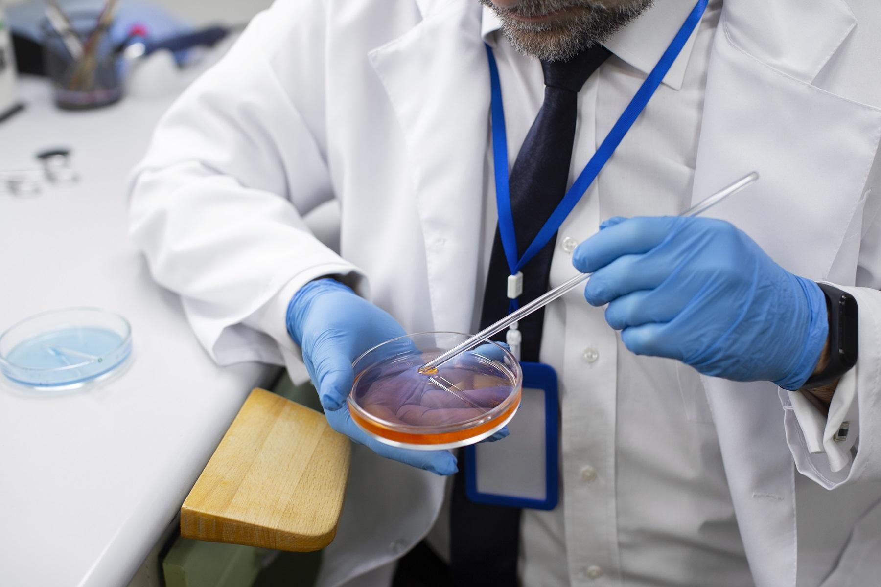
A histopathology - radical resection specimen test is a detailed laboratory examination of tissue removed during cancer surgery. The surgeon removes the tumour along with a surrounding border of healthy tissue and nearby lymph nodes, and this entire specimen is then sent to a pathology laboratory for analysis.
Also known as histopathology – large complex / cancer specimen or biopsy material radical, the test helps doctors understand the exact nature of the disease. Results guide decisions about further treatment after surgery.
What Does a Histopathology - Radical Resection Specimen (Histopath (Radical)) Test Measure?
This test is a detailed examination of multiple features of the removed tissue. Rather than producing numerical readings, it generates a descriptive report covering several key aspects of the specimen. The following parameters are assessed during the examination:
| Parameter | What It Tells the Doctor |
|---|---|
| Tumour type | Whether cells are malignant, and which type of cancer is present |
| Tumour grade | How abnormal the cells look compared to normal cells (Grade 1 = well differentiated; Grade 3 to 4 = poorly differentiated) |
| Tumour size and extent | Dimensions of the primary tumour and how deeply it has invaded surrounding tissue |
| Resection margins | Whether cancer cells are present at the cut edges of the removed tissue (positive = cells present; negative = cells absent) |
| Lymph node status | Whether examined lymph nodes contain cancer cells (positive) or do not (negative) |
| Lymphovascular invasion | Presence of cancer cells inside blood vessels or lymphatic channels |
| Perineural invasion | Whether cancer cells are found around or along nerve fibres |
| TNM staging | Pathological staging of the primary tumour (pT) and regional lymph nodes (pN) according to international guidelines |
Why Is Is a Histopathology - Radical Resection Specimen (Histopath (Radical)) Test Done?
This test is ordered after a radical surgical procedure to fully characterise the removed tissue and guide the next steps in a patient's care.
Common Symptoms That May Require This Test
A doctor may recommend surgery followed by a histopathology large specimen (with lymph nodes) examination when a patient presents with the following:
- An unexplained lump or mass that has been identified on examination or imaging
- Persistent pain in a localised area without a clear cause
- Unexplained or significant weight loss over a short period
- Abnormal bleeding not explained by other conditions
- Noticeable changes in the function of an organ
- Suspicious or inconclusive findings on an X-ray, CT scan, or MRI
Conditions This Test Can Help Detect
The histopathology - radical resection specimen test can provide diagnostic clarity for several conditions, including:
- Malignant cancers of various organs, including the colon, prostate, breast, bladder, and uterus
- Benign or malignant tumours requiring full characterisation
- Inflammatory bowel conditions such as ulcerative colitis or Crohn's disease when surgical resection has been performed
- Uterine fibroids examined after surgical removal
- Assessment of how completely a tumour has been surgically removed
- Evaluation of the tissue's response to chemotherapy or radiotherapy given before surgery
How to Prepare and What to Expect
Preparation for this test centres on the surgical procedure itself rather than the laboratory analysis. Your surgical team will provide specific pre-operative instructions.
Do You Need to Fast?
No fasting is required for the laboratory analysis of the specimen. However, fasting is typically required before the surgical procedure used to collect the specimen. Follow your surgeon's instructions precisely regarding food and fluids before the operation.
Practical Tips Before Your Test
Keep the following points in mind in the lead-up to your procedure:
- Inform your doctor of all medications you take, including supplements and over-the-counter drugs, as some may need to be paused before surgery
- Disclose any known allergies, particularly any past reactions to anaesthesia
- Share your complete medical history, previous biopsy reports, and recent imaging results with your doctor
- Arrange for a family member or friend to accompany you and drive you home after the procedure if general or regional anaesthesia is used
Step-by-Step Procedure
- You will be given anaesthesia (local, regional, or general) as decided by your surgical team before the procedure begins.
- The surgeon removes the tumour together with a border of surrounding tissue and the relevant lymph nodes as a single specimen.
- The specimen is immediately placed in a specialised fixative solution by the surgical team within minutes of removal to preserve the tissue structure accurately for analysis.
- In the laboratory, the tissue is processed, embedded in paraffin wax, and cut into very thin sections for staining.
- A qualified pathologist examines the sections under a microscope, assessing all key parameters including margins, grade, and lymph node status.
- The completed pathology report is prepared and made available to your doctor within the stated turnaround time.
Factors That Can Affect Accuracy
The following factors can influence the quality and accuracy of the final report:
- Delayed or improper fixation of the specimen after removal
- Incomplete clinical information provided to the pathologist
- Inadequate orientation or inking of surgical margins before processing
- Insufficient tissue sampling from large or complex specimens
- Need for additional tests such as immunohistochemistry, which can extend the reporting time
Understanding Your Histopathology - Radical Resection Specimen (Histopath (Radical)) Test Results
Results from this test are descriptive rather than numerical. Your doctor will review the full report together with your imaging findings and clinical history to plan your next steps.
The table below explains the key findings you may see in your report:
| Parameter | Favourable Finding | Concerning Finding |
|---|---|---|
| Margins | Negative (clean): no cancer cells at cut edge | Positive (involved): cancer cells present at edge |
| Lymph nodes | Negative: no cancer cells detected | Positive: cancer cells present in one or more nodes |
| Tumour grade | Grade 1: cells closely resemble normal tissue | Grade 3 to 4: cells appear highly abnormal |
| TNM Stage | pT1 to pT2, pN0, cM0 (localised disease) | Higher T, N, or M values indicate wider spread |
| Lymphovascular invasion | Absent | Present |
| Perineural invasion | Absent | Present |
These ranges are general guidelines. Your doctor will interpret your results based on your age, health history, and other factors. Always consult a qualified healthcare professional for personalised medical advice.
Managing Your Recovery and Post-Surgical Care
While this test examines surgically removed tissue rather than ongoing blood values, the following general points support recovery and follow-up care:
- Attend all scheduled post-surgical follow-up appointments and imaging reviews as advised by your oncology team
- Eat a balanced diet with adequate protein, fruits, and vegetables to support healing after surgery
- Avoid smoking and limit alcohol, as these can affect recovery and overall health outcomes
Lupin Diagnostics Histopathology - Radical Resection Specimen (Histopath (Radical)) Test Price
Home sample collection is not available for surgically resected tissue specimens. Whole surgical specimens must be submitted directly to a designated Lupin processing hub via authorised hospital logistics or clinical hand-delivery by a representative.
The approximate histopathology - radical resection specimen test price across major Indian cities is listed below.
| City | Approximate Price (₹) |
|---|---|
| Mumbai | ₹3,000 |
| Bangalore | ₹3,000 |
| Chennai | ₹3,000 |
| Hyderabad | ₹3,000 |
| Kolkata | ₹3,000 |
| Indore | ₹3,000 |
| Pune | ₹3,000 |
| Bhopal | ₹3,000 |
Prices are indicative and may vary by location. Please confirm the current price at the time of booking.
How to Book
- Your hospital surgical team will remove the radical specimen during your scheduled surgery
- Ensure the hospital staff immediately places the whole organ or tumour specimen into a 10% buffered formalin container to preserve the tissue
- Select the test on the Lupin Diagnostics website.
- Contact Lupin Diagnostics customer support or coordinate with your hospital's laboratory management team to initiate a professional sample handover
- Authorised medical courier teams or clinical representatives will transport the preserved specimen directly to your city's preferred Lupin processing hub
- Receive your report via email or WhatsApp within the stipulated turnaround time
Frequently Asked Questions
A histopathology - radical resection specimen is tissue removed during a major cancer surgery. It includes the tumour itself, a surrounding margin of healthy tissue, and nearby lymph nodes. The goal of the laboratory examination is to determine how far the disease has spread and whether the surgical removal was complete.
For most cases, the report is ready within 5 to 10 working days. If the pathologist needs to perform additional tests such as immunohistochemistry or special stains, the turnaround can extend to 7 to 14 days. Your doctor will let you know if additional testing is required.
Lymph nodes close to the tumour may contain cancer cells that have spread from the primary site. The pathologist examines these nodes to determine whether the cancer has moved beyond its original location. This information is essential for accurate staging and for deciding whether additional treatment such as chemotherapy is needed.
Negative margins mean the pathologist found no cancer cells at the outer edges of the removed tissue. This suggests the surgeon was able to remove all visible cancer. Your doctor will explain whether any further treatment is still recommended based on the full report.
No. The tissue specimen for this test is obtained during a surgical procedure in a hospital or clinical setting under a doctor's supervision. It cannot be collected at home. You will need to have the specimen submitted directly from the surgical facility via authorised hospital logistics or clinical handover.
Immunohistochemistry (IHC) is an additional laboratory technique that identifies specific proteins or markers in cancer cells. It is sometimes required to confirm the type of cancer or to identify targets for specific treatments. When IHC is needed, it adds to the overall turnaround time for the report.
Yes. The pathology report from a histopathology - large complex / cancer specimen examination includes pathological TNM staging, which describes the size and extent of the tumour, the involvement of nearby lymph nodes, and any evidence of spread. Your oncologist will use this staging information alongside other clinical findings to plan your treatment.
Histopathology Radical Resection Specimen Test: Purpose, Procedure, Results & Price